Click the image to continue
When a doctor needs to see inside your body, they may order an imaging test. The names can sound similar, but each scan works differently and is better for certain problems. Knowing the basics helps you feel calmer and prepare properly.
X-Ray
An X-ray uses a small dose of radiation to create a picture, mostly of bones and the chest. It is quick, widely available and inexpensive. X-rays are commonly used for fractures, chest infections, joint problems and dental checks. The procedure takes only a few minutes and needs little or no preparation, although you may be asked to remove jewellery and metal objects.
CT Scan (Computed Tomography)
A CT scan takes many X-ray images from different angles and a computer combines them into detailed cross-sectional pictures. It is excellent for head injuries, internal bleeding, lung problems, kidney stones and many emergencies because it is fast. A CT scan uses more radiation than a plain X-ray, so doctors order it when the benefit is clear. Some CT scans use a contrast dye given by mouth or injection; you may be asked about allergies and kidney function beforehand.
MRI (Magnetic Resonance Imaging)
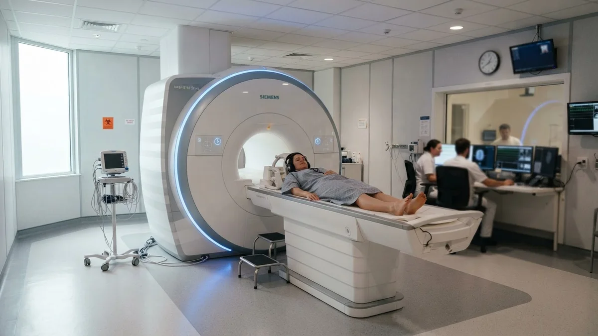
MRI uses a strong magnet and radio waves instead of radiation. It produces very detailed images of soft tissues such as the brain, spinal cord, ligaments, muscles and some organs. An MRI takes longer, often 20 to 60 minutes, and the machine is noisy, so earplugs or headphones are provided. Because of the magnet, you must tell staff about pacemakers, metal implants, clips or any metal fragments in the body. People who feel uncomfortable in enclosed spaces should mention it in advance.
Ultrasound
Ultrasound uses sound waves and a handheld probe with gel on the skin. It has no radiation, so it is widely used in pregnancy and for checking the abdomen, thyroid, blood vessels, heart (echocardiography) and soft-tissue lumps. Some abdominal scans need fasting, and pelvic scans may need a full bladder. Ultrasound is painless and usually takes 15 to 30 minutes.
Comparison at a Glance
| Test | Uses radiation? | Typical time | Best for |
|---|---|---|---|
| X-ray | Yes, low dose | Few minutes | Bones, chest |
| CT | Yes | 5–15 minutes | Emergencies, detailed body images |
| MRI | No | 20–60 minutes | Brain, spine, joints, soft tissue |
| Ultrasound | No | 15–30 minutes | Pregnancy, abdomen, vessels |
How to Prepare
- Read the preparation sheet given by the radiology department.
- Tell staff if you are or might be pregnant.
- Mention allergies, kidney problems, diabetes medicines and implants.
- Wear comfortable clothes without metal zips or buttons.
- Bring previous scans and reports for comparison.
Getting the Results
A radiologist, a doctor trained to read scans, studies the images and writes a report. Your own doctor then explains what it means for you. Ask for a copy of the report and images for your records, as they are useful for future comparisons.
Conclusion
Each imaging test has its strengths. Your doctor chooses the one that gives the most useful information with the least risk. Asking questions and following preparation steps helps the scan go smoothly.
Disclaimer: This article is for general information only and is not a substitute for professional medical advice. Always consult a qualified healthcare provider about your own situation.